The Use of Neuroimaging in the Study of Mental Disorders
Mental health was long considered to be something invisible. Physicians and psychiatrists had to completely depend on what an individual said or how he or she behaved to determine what was amiss. In case a person was deeply sad, or constantly feared something, there was no physical examination, say a blood test or an X-ray to prove it. This meant that many people could not easily feel comprehended and tended to feel shame that their plight was not real since nobody could see it on a screen. The modern science has however altered the game. We are leaving behind the discussion of symptoms and entering the area of biological map of the brain.
Making the Invisible Visible
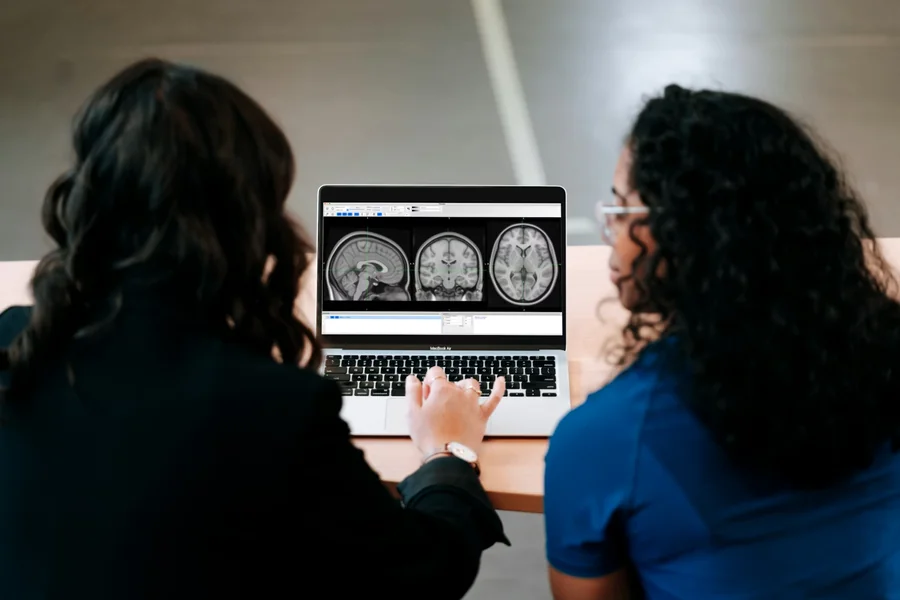
The most significant change in the contemporary psychology is the possibility of visualizing the brain at work. There is no longer the need to speculate about what is going on behind the scenes. Scanning the brain, which is also known as neuroimaging, enables the researchers to see the physical alterations that occur when an individual is experiencing the problem of the mental disorder. This shift is comparable to the one in which we are using a paper map and then the advanced GPS, it allows us to have a much better picture of the locations of the traffic jams in the mind. You can explore more about how these scans are changing the way we treat patients over at the Liven mental health blog, where we bridge the gap between complex neuroscience and daily well-being. By turning an invisible struggle into a visible reality, these scans prove that mental health is physical health.
The Different Ways We Look at the Brain
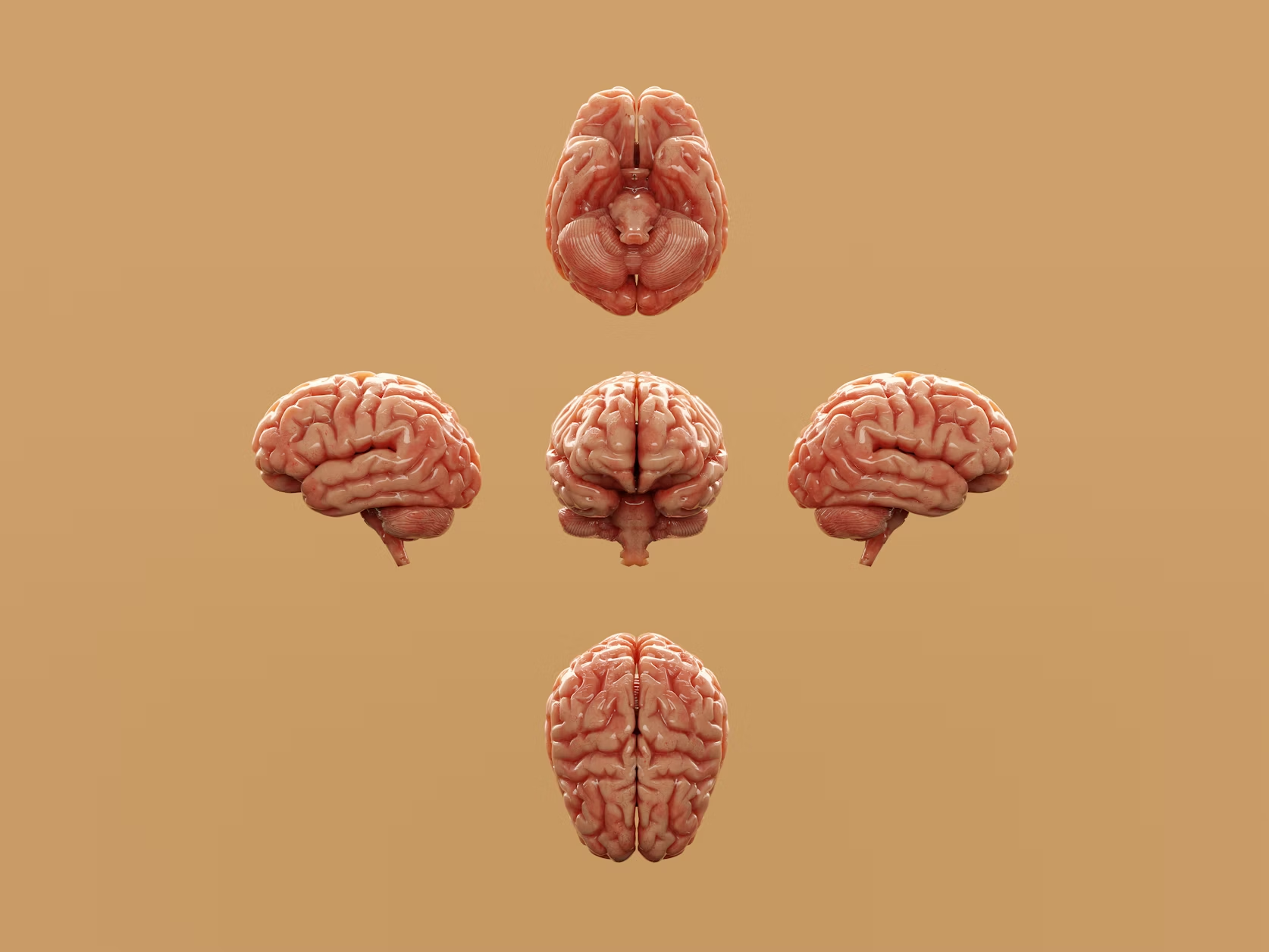
It is not only possible to take a picture of the brain in a single way. There are three principal tools utilized by scientists, and each has a specific task. The first is the Structural MRI. It is as though you were making a high-definition photograph of a building to examine its bones. It aids physicians in examining whether some sections of the brain are actually smaller or slimmer than what they ought to be. Second, there is the Functional MRI (fMMRI). This device is an actual tracking of the flowing blood in real-time, as opposed to merely viewing the form. It tells us which areas of the brain are becoming over-active, such as a car with an overheating engine, and which areas of the brain are remaining under-active. Lastly, PET scans, or as they are referred to as the mailmen of the brain or neurotransmitters of the brain, are also involved. These scans are used to map chemicals such as dopamine and serotonin to determine whether the brain communication system is passing the message in the right way.
Why Some People Can’t Feel Joy
Brain scans are revolutionizing our knowledge on anhedonia which is inability to feel joy. Researchers have used functional MRI to realize that the brain reward system, in many cases, is not active in depressed people. In particular, the ventral striatum, which is one of the central components of the pleasure center, does not light up when provoked by activities previously appreciated.
Such scans also indicate a physical disconnectivity of the emotional and logical brain areas with each other not communicating effectively. This objective data is a breakthrough in the clinical care. Doctors are able to go beyond guesswork by visibly experiencing the point of the neural communication failure. They can establish whether a patient requires special drug to stimulate the reward circuitry or special talk therapy to reconnect such crucial connections in the brain.
Mapping Out Fear and Stress
The brain scans of the anxiety and PTSD show an over-reacting alarm system. The amygdala is a small almond-shaped panic button that can tend to be bigger or more reactive in nervous people, which releases whenever there is an indication of trouble, and therefore, the body is already on high alert.
The hippocampus, the memory vault of the brain of those with PTSD, is physically altered by trauma. This area tends to store experiences as the past occurrences, yet when traumatized, it cannot store the terrifying memories in the right place. This makes the past to repeat itself in the present. The brain practically becomes uncontrollable and the logic center finds it hard to suppress the fear response once it sets in.
Seeing How the Brain Grows and Changes
Schizophrenia can be investigated using neuroimaging, which can demonstrate that there are physical variations in the brain structure, which frequently include the enlargement of the fluid-filled spaces as the brain tissue becomes thin. As functions, the frontal lobes, the planning and organizing centers, often can be used in a low power mode, the part of the brain that cannot meet the energy requirements of intricate activities.
The scans of the brain of the present day also monitor cables or neural links. In other instances, these pathways either get fragmented or rather woven during the crucial teenage years. This discovery of such wiring problems enables scientists to determine where and when to interfere. Clinicians can support the development of the brain of a young person more effectively by visualizing these biological shifts, which will be moving towards earlier and more accurate treatment of complicated mental health issues.
The Future of Brain Scans
It is a new age of brain scans that could be used to make early warnings. Similarly to the example of the heart scan that reveals a blockage in the heart before a heart attack, brain scans may one day reveal signs of being at-risk before someone even starts experiencing a change in his or her mental health. Also, there is a new exciting branch known as the neurofeedback. In this treatment, a patient can view his or her brain in real-time on a screen. The ability to train their brain to make it discover its own balance is the actual result of seeing what thoughts or breathing exercises are calming their overactive amygdala. Naturally, this technology raises the issues of privacy on a big scale. Having a literal map of your own mental health is a potent thing and we need to make sure that these data are applied in an ethical way to help people instead of categorizing them.
Summing Up
It is a new frontier in the discovery of self to shift the line of questioning the other person on what they are doing, to what your brain is doing. Neuroimaging breaks down the stigma and shame that accompanies mental health by exposing the physical causes of mental disorders. Although these scans are not a kind of magic crystal ball, they can be used as the guide through the mind. When the disorder is observed on a screen it confirms to the millions of people that they are not alone in their suffering and that their brains are just not wired the same way. Visualizing this neural map gets us nearer to the era when the field of mental health care will be just as accurate and transparent as the field of medicine.